[P-027]
Kalsifiye spermatik kord kisti: Olgu sunumu
Hüseyin Koçan1, Şiir Yıldırım2, Mehmet Yazıcı3, Erhan Erdoğan1, Enver Özdemir12Kanuni Sultan Süleyman Eğitim ve Araştırma Hastanesi Histoloji ve Embriyoloji Kliniği, İstanbul
3İstanbul Eğitim ve Araştırma Hastanesi Çocuk Cerrahisi Kliniği, İstanbul
Dejenerasyon ya da nekroz gibi doku değişikliklerinin üzerine kalsiyum tuzlarının birikmesine distrofik kalsifikasyon denir. Vücudun herhangi bir yerinde oluşabilmekle birlikte, en sık akciğer tüberkülozuna sekonder gelişmekte olup, ürogenital sistemde oldukça nadir görülmektedir. Tüberkülozda kazeifikasyon nekrozu alanlarında, ölmüş ve kapsüllenmiş parazit nodüllerinde, hidatik kistlerde kist etrafında, eskimiş tromboz ve nedbelerde, aktinomikoz, botriyomikoz ve stafilokok granulomlarında, eski apselerde, Zenker nekrozunda, bazı benign ve malign tümörlerde distrofik kalsifikasyon meydana gelebilir. Funikulus spermatikusta benign lezyonlar ile tüm yaş gruplarında sık karşılaşılmasına karşın, cerrahi hastalıklar en sık çocuk yaş grubunda saptanmakta ve tüm yaş gruplarında kalsifiye kitleler oldukça nadir görülmektedir. Biz de burada, inguinal bölge yerleşimli 4 cm boyutunda sert nodüler kitle ile tarafımıza başvuran 44 yaşındaki bir hastayı sunmayı amaçladık.
Calcified spermatic cord cyst: a case report
Hüseyin Koçan1, Şiir Yıldırım2, Mehmet Yazıcı3, Erhan Erdoğan1, Enver Özdemir12Department of Histology and Embryology, Kanuni Sultan Suleyman Training and Research Hospital, Istanbul, Turkey
3Department of Pediatric Surgery, Istanbul Training and Research Hospital, Istanbul, Turkey
Dystrophic calcification is the accumulation of calcium salts in the tissue alterations such as degeneration or necrosis. Although it may occur on any part of the body; it develops most often secondary to pulmonary tuberculosis and extremely rare in the urogenital tract. Dystrophic calcification can occur in the areas of caseation necrosis in tuberculosis, in dead and encapsulated parasitic nodules, around the cysts in hydatid cysts, in obsolete thrombosis and scars, in the granulomas of actinomycosis, botryomycosis and staphylococcus, in old abscess, in Zenker’s necrosis, in some benign and malignant tumors. Alltough the benign lesions of funiculus spermaticus are encountered in all age groups; the surgical diseases are detected most often during childhood and calcified masses are quite rare in all age groups. We aimed to present a 44-year-old patient who was admitted us with solid nodular mass 4 cm in size located in the inguinal region with the review of the literature.
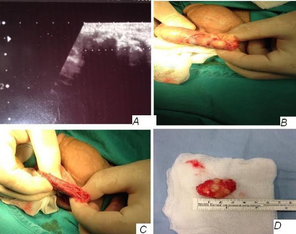
Resim 1A
Picture 1A
)
Resim 1A: Ultrason görüntüsü, B: İnguinal eksplorasyon C: Eksizyon sonrası spermatik kordun görüntüsü, D: Makroskobik görüntüsü
Picture 1:A Ultrasound image, B: Inguinal exploration C: After excision of the spermatic cord image, D: Macroscopic image