[P-025]
Kistik Bening Prostat Hiperplazisi
İbrahim Karabulut1, Ercüment Keskin2, Bakyt Kozubaev6, Oğuz Demirdöğen6, Fevzi Bedir1, Erdem Koç3, Ali Haydar Yılmaz4, Mahmut Koç4, Hüseyin Koçakgöl5, Fatih Özkaya1, Fatih Kürşat Yılmazel12Mengücek Gazi Üniversitesi Eğitim ve Araştırma Hastanesi Erzincan
3Yıldırım Beyazıt Üniversitesi Yenimahalle Eğitim ve Araştırma Hastanesi Ankara
4Bilecik Devlet Hastanesi Bilecik
5Kanuni Eğitim Ve Araştırma Hastanesi Trabzon
6Atatürk Ünversitesi Tıp Fakültesi Üroloji ABD Erzrurum
GİRİŞ:
Prostatta görülen tartışılan kistik lezyonlar mülleriyan kanal kisti, ejakülatör kanal kisti, benign prostat hiperplazisi kistik dejenerasyon, prostat tutan kistler, kaviter prostatit ve prostat apsesidir. Bunlar müllerin kanal kistlerive utrikul olarak katagorize edilebilir.
Prostat kistlerinin çoğu asemptomatiktir ve %5 oranında az bir kısmı semptomatiktir. Semptomatik olan hastalarda hemotaspremi, işeme disfonksiyonları ve prostatit benzeri şikayetler gözlenebilir.
Literatürede tedavisinde ince iğne aspirasyonu, transüretral rezeksiyon(TUR) hatta açık cerrahi tarif edilmekle beraber önerilen yöntem TUR yaklaşımdır. Tek odakli ve büyük kistlerde ince iğne aspirasyon yöntemi denenebilir.
Biz bu olgumuzda yaopılan tetikleri esnasında prostatik loj mesane taban yerleşimli multipl septalı kist tespit edilen hastayı sunmayı ve literatür bilgilerini gözden geçirmeyi amaçladık.
OLGU:
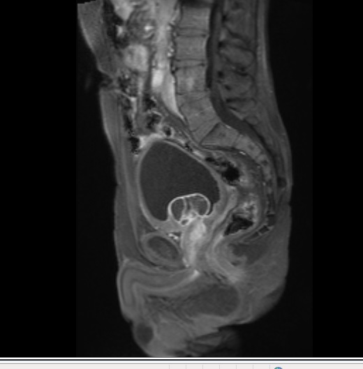
Elli üç yaşında erkek hasta sık sık idrara çıkma, idrar yaparken zorlanma ve gece idrara kalkma şikayeti nedeni ile polkliniğimize başvurdu.. Yapılan fizik muayenede patoloji tespit edilmedi. Rektal tuşede prostat grade II cesaemtte doğal idi. İstenilen laboratuar tetikleri normal idi. Yapılan üroflowmetride maksimum akın hızı: 11ml/sn, post voiding rezidü: 65 cc idi. Poliklinik şartlarında yapılan ultrasonografide prostatik lojda kistik görünüm tespit edilmesi üzerine hastadan alt batın pelvik manyetik rezonans (MR) istendi. Rapor sonucunda; mesane tabanında yaklaşık 4.5x4,5 cm ebatlı, T1AG’ de hipointens, T2AG’de hiperintens multpl septasyonlar içeren, postkontrast serilerde septal ve duvar kontrastlanması gösteren lezyon alanları(Resim 1,2) izlendi. Görünümün prostatın kistik adenom veya kistik adenokarsinomu ile uyumlu olabileceği raporlandı. Kist hidatik serolojisi negatif olarak değerlendirildi. Lezyon odaklı olmak üzere 12 kadran prostat biopsisi alındı. Patoloji sonucu bening prostat hiperplazisi(BPH) ile uyumlu olarak tespit edildi. Prostatism semptomlarının ön plan olması nedeni ile hastaya endoskopi yapıldı. Sistoskopide median lop hiperplazisi izlendi. Mesanede tarbekülasyon artışı izlendi. Lezyon sahası olduğu düşünülen median lopa transüretral rezeksiyon yapıldı. Hasta post operatif üçüncü gün komplikasyonsuz olarak taburcu edildi. Patoloji sonucu BPH ile uyumlu olan hastaya çekilen MR’da(Resim3) patoloji tespit edilmedi. Hasta poliklinik kontrollerine gelmektedir.
SONUÇ:
Prostat kistleri değişik görünüm(radyolojik) ve aynı ölçüde farklı semptomlarla başvurabilmektedir. Tedavi planını belirlerken hastanın öncelikli semptomları, yaşı, kistin boyutu, tek veya multipl oluşu, natürü ve serum PSA değerleri göz önünde bulundurulmalıdır. Ancak tedavi ve takip protokollerini belirlemede daha geniş hasta serilerini içeren çalışmalara ihtiyaç vardır.
Cystic Benign Prostatic Hyperplasia
İbrahim Karabulut1, Ercüment Keskin2, Bakyt Kozubaev6, Oğuz Demirdöğen6, Fevzi Bedir1, Erdem Koç3, Ali Haydar Yılmaz4, Mahmut Koç4, Hüseyin Koçakgöl5, Fatih Özkaya1, Fatih Kürşat Yılmazel12Mengücek Gazi University Education and Research Hospital Erzincan
3Yildirim Beyazit University Yenimahalle Training and Research Hospital Ankara
4Bilecik State Hsospital Bilecik
5Kanuni Education and Resarch Hospital Trabzon
6Atatürk Universty Medicine Faculty Urology Service Erzurum
INTRODUCTION:
The debated cystic lesions that are common in prostate include mullerian duct cyst, ejaculatory duct cyst, benign prostatichyperplasia cystic degeneration, cysts that involve prostate, cavitary prostatitis and prostatic abscess. They can be categorized into müller’s duct cysts and utricle.
We present a case with a cyst with multiple septas localized in the prostatic area on the bladder bottom detected during tests, and reviewed the literature.
CASE:
A 53-year-old male patient admitted our polyclinics for thamuria, difficulty in urination, and nocturia. The physical examination revealed no pathologies. Rectal palpation indicated prostate grade II and the size was normal. The ordered laboratory tests were normal. The uroflowmetry showed that maximum flow rate: 11ml/s, post voiding residue: 65 cc. The ultrasonography taken under polyclinic conditions demonstrated a cystic appearance in the prostatic area, therefore magnetic resonance of lower abdominal pelvic was performed on the patient. The report results showed areas of lesion of 4.5x4.5 cm with septal and wall contrast in post-contrast series containing multiple hypo-intense septations in T1AG and hyper-intense septations in T2AG on the bladder bottom (Figures 1,2). The appearance was reported to be consistent with cystic adenoma or cystic adenocarcinoma of prostate. The hydaticserology of the cyst was negative. A prostatic biopsy of 12 quadrants was performed, focusing on the lesion. The results of the pathology were consistent with the benign prostatic hyperplasia (BPH). An endoscopy was performed on the patient due to apparent symptoms of prostatism. The cystoscopy revealed hyperplasia of median lobe. The bladder had increased trabekülasyon. The patient was discharged at postoperative day 3 without complications. The results of the pathology were consistent with BPH, therefore an MR was taken (Figure 3) which showed no pathology. The patient still visits the polyclinic for routine checks.
RESULTS
Prostatic cysts may have different appearances (radiologic) and different symptoms of same size. The patient’s symptoms, age, the size of the cyst, one or multiple cysts, the nature of the cyst, and serum PSA values should be considered for the treatment plan. However, studies including a large series of patients are needed to decide on the treatment and follow-up protocols.
Resim 1
Picture 1
)
Resim 2
Picture 2

)
Resim 3
Picture 3

)