[P-244]
Oturum adı: POSTER SESSION 4 | Oturum salonu: POSTER AREA | Oturum tarihi: 18 Ekim 2014 | Oturum saati: 08:00 - 13:00Nadir Görülen Benign Bir Testis Tümörü: Epidermoid Kist Olgu Sunumu
Tolga Şahin, Cihat Özcan, Ersin Atabey, Çağrı Şenocak, Ömer Faruk BozkurtGİRİŞ: Testiküler epidermoid kistler benign epitelial tümörler olup tüm intratestiküler kitlelerin %1 ‘ini oluşturur. Genellikle testiste ağrısız palpable kitle mevcuttur ve tümör markerları normal sınırlardadır. Tipik olarak küresel lezyonlardır. Histolojik olarak fibröz bir kapsülü olan gerçek kistlerdir. Kist içeriğinde ya da duvarında kalsifikasyonlar içerebilir. Ultrasonografik bulguları spesifik olmamakla birlikte sonografide soğan kabuğu manzarası ve kanlanma bulunmaması önemlidir ve epidermoid kisti düşündürmelidir. Bütün vakalarda eksplorasyon gereklidir.
OLGU: Ağrısız sol testiküler şişlik şikayetiyle başvuran, 21 yaşındaki erkek hastanın muayenesinde sol testis ortasında düzgün sınırlı, yaklaşık 10x10 mm' lik sert kitle mevcuttu. Tümör markerları ve diğer laboratuar sonuçları normaldi. Ultrasonda sol testis orta kesimde 13x12 mm, santralinde 3 mm kalsifikasyon içeren heterojen hipoekoik solid düzgün sınırlı lezyon mevcuttu. Doppler incelemede belirgin kanlanma izlenmedi. Bulgular ışığında benign bir lezyon olabileceği düşünülerek testis koruyucu cerrahi planlandı. Sol inguinal kesi ile testis eksplore edildi. T. albuginea açılarak testis ortasındaki 10x10 mm' lik düzgün sınırlı sert kitle lezyonu eksize edildi. Enükle edilen kitleden ve tabandan ayrı ayrı frozen gönderildi. Enükle edilen lezyon epidermoid kist, taban örneklemesi normal testis dokusu gelmesi üzerine işleme son verildi. Nihai patolojide ise çok katlı epiteli bulunan ve atipi gözlenmeyen epidermoid kist izlendi. Hastanın 3 yıllık takiplerinde nüks ya da metastaz izlenmedi.
SONUÇ: Epidermoid kist için testis koruyucu cerrahi benign bir lezyon olduğunu gösteren bulguların varlığında mümkün olabilir: Bunlar tipik ultrasonografi bulguları(soğan kabuğu görünümü,kanlanma olmaması), tümör markerlarının normal olması, 2 cm’den küçük olması,tanının frozen incelemeyle doğrulanmasıdır.
A Rare Benign Testis Tumour: Epidermoid Cyst Case Report
Tolga Şahin, Cihat Özcan, Ersin Atabey, Çağrı Şenocak, Ömer Faruk BozkurtINTRODUCTION: The epidermoid cyst of the testis is a rare benign tumour. It accounts for approximately 1% of all testicular tumours. Generally, the testis has a palpable, painless mass with tumour markers within normal ranges. They are typically spheric lesions. Histologically it is a real cyst which has a fibrous capsule. Calsifications may be demonstrated in the cyst content or cyst wall. Although ultrasonografic findings are non-spesific, absence of vascular flow and onion-like appearance are important and call to mind the diagnosis.Exploration is necessary for all patients.
CASE: A 21-year-old male visited our outpatient department complained having a painless hard mass in the left testis for about one week. Physical examination showed a hard mass measuring 10x10 mm was palpable in the center of the left testis. Tumour markers and other laboratory results were normal. An ultrasound examination demonstrated a 13x12 mm, well-circumscribed, heterogeneous, hipoechoic, round, solid mass in the center of the left testis with a 3 mm calcification area in it. Absence of vascular flow was also noted on colour Doppler sonography. Under the impression of a benign lesion, testis-sparing surgery was arranged with inguinal incision and frozen section biopsy of the left testicular tumour did not show any evidence of malignancy, hence enucleation of the tumor was performed. Final pathology was reported that a epidermoid cyst was lined by squamous epithelium without any atypia. At 3-year follow-up the patient had no relapse or metastasis.
CONCLUSIONS: Testis-sparing surgery might be possible if there is significant suspicion of a benign lesion: typical sonographic findings(onion-like appearance, absence of vascular flow), normal tumour markers, size less than 2 cm, frozen section histological examination confirming the diagnosis.
resim-1
picture-1
)
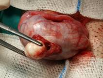
Lezyonun intraoperatif görünümü
intraoperative appearance of the lesion
resim-2
picture-2
)
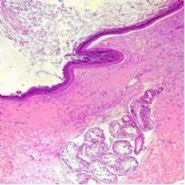
Kisti çevreleyen çok katlı keratinize epitelin bulunduğu histopatolojik görüntü X20, hemotoksilen eozin boyama ile
Histopathology at 20X with hematoxilen and eosin stain demonstrates squamous epithelium of the cyst
resim-3
picture-3
)
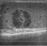
epidermoid kistin tipik USG bulgusu,soğan kabuğu görünümü
typical ultrasonografic finding of epidermoid cyst,onion-like appearance