[P-236]
Oturum adı: POSTER SESSION 4 | Oturum salonu: POSTER AREA | Oturum tarihi: 18 Ekim 2014 | Oturum saati: 08:00 - 13:00Skrotal Leiyomyom
Şahin Kılıç, Yusuf Gençten, Engin Kölükçü, Fikret ErdemirAMAÇ: Leiyomyomlar iyi huylu tümörlerdir ve düz kas içeren herhangi bir yapıdan veya organdan ortaya çıkabilirler. Genellikle uterusta (% 95) görülmelerine rağmen, ürogenital sistemde renal pelvis, mesane, spermatik kord, epididim, testis, prostat, glans penis ve skrotumda da bildirilmişlerdir. Tunika dartostan köken alan skrotal cildin leiyomyomları son derece nadir görülen benign tümörlerdir. Bu olguda amacımız skrotum cildinde kitle ile kliniğe başvuran skrotal leiyomyom olgusunu sunmak.
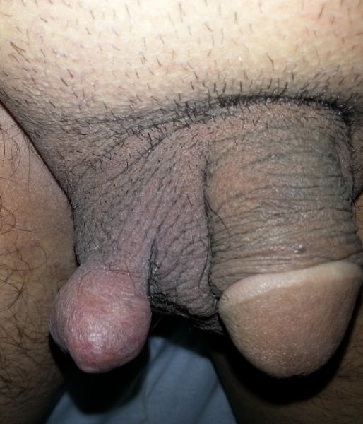
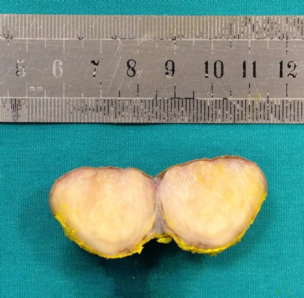
OLGU: Elli yaşında erkek hasta skrotum cildinde kitle ile kliniğimize başvurdu. Öyküsünde özellik yoktu. Fizik muayenede skrotum sağ ön yüzde yaklaşık 3x4 cm ebatlarında düzgün sınırlı, orta sertlikte, yuvarlak, mobil kitle gözlendi.[resim1] Hassasiyet ve sistemik bulgu saptanmadı. Laboratuar tetkikleri normaldi. USG de kitle solid, heterojen ve hipoekoik olarak izlendi. Lokal anestezi ile çapı 3 cm, yuvarlak şekilli, sarı renkli, kapsülsüz solid nodüler kitle eksize edildi.[resim 2] Histomorfolojik ve immünhistokimyasal bulgular ışığında skrotal cilt leiyomyomu olarak değerlendirildi. Altı ay takip sonrası nüks izlenmedi.
SONUÇ: Sonuç olarak skrotal leiyomyomun nadir görülmesine rağmen, skrotum cildinde solid kitle ile başvuran hastalarda ayırıcı tanıda göz önünde bulundurulması gerektiğini düşünüyoruz.
Scrotal Leiomyoma
Şahin Kılıç, Yusuf Gençten, Engin Kölükçü, Fikret ErdemirAIM: Leiomyomas are benign neoplasms and they may arise from any structure or organ containing smooth muscle. Although they are often seen in the uterus (95%), in urinary system in the renal pelvis, bladder, spermatic cord, epididymis, testis, prostate, the glans penis or scrotum have been reported. Genital leiomyomas of the scrotal skin are extremely rare benign tumors, originating from the tunica dartos of the scrotum. In this case our aim is to present a case of scrotal leiomyoma, admitted to the clinic with a mass on the scrotum.
CASE: A 50- year-old man admitted to our clinic with a scrotal mass. His history was unremarkable. On physical examination a 3x4 cm in diamater well defined, medium hardness, round, mobile regular mass in the right scrotal area [Picture 1] was detected. We did not find the tenderness and systemic examination was unremarkable. Biochemical and routine hematologic analysis were within normal range. Ultrasonography demonstrated a solid heterogenous, hypoechoic mass. With local anesthesia, 3 cm in diameter, round-shaped, yellow-colored, encapsulated solid nodular mass [Picture 2] was excised. The tumoral lesion was evaluated as leiomyoma of scrotal skin on the basis of histomorphological and immunohistochemical findings. Six months after surgery, there were no recurrence.
CONCLUSION: We think that, although leiomyoma of the scrotum is rare, it should be considered in the differential diagnosis in patients presenting with solid mass on the scrotum.
resim 1
picture 1
)
resim 2
picture 2
)