[P-194]
Oturum adı: POSTER SESSION 4 | Oturum salonu: POSTER AREA | Oturum tarihi: 18 Ekim 2014 | Oturum saati: 08:00 - 13:00Erişkin Hastada Agresif Seyirli Wilms Tümörü Olgusu
Emre Altıntaş1, Mustafa Kucur1, Pınar Karabağlı2, Murat Gül1, Mehmet Kaynar1, Serdar Göktaş12Selçuk Üniversitesi Tıp Fakültesi, Patoloji Ana Bilim Dalı, Konya
Amaç
Wilms' tümörü çocuklarda en sık görülen primer malign böbrek tümörüdür. Erişkinlerde ise oldukça nadir görülmektedir. Erişkin hastada agresif seyirli wilms tümörünün tanı ve tedavisi sunulmuştur
Method ve Olgu
28 yaşında erkek hastanın 2 aydır sol yan ağrısı ve hematüri nedeniyle dış merkezde yapılan değerlendirilme sonrası çekilen ultrasonografide sol böbrekte yaklaşık 10x8 cm kitle saptanması üzerine kliniğimize refere edilmiştir. Hastada yapılan fizik muayene ve rutin laboratuvar tetkikleri normal olarak saptandı. MR görüntülemede sol böbrek üst-orta pol lokalizasyonunda 10x8 cm boyutunda içinde kistik komponenti bulunan ve nekroze alanlar içeren, belirgin kontrast tutulumu gösteren T1 ağırlıklı görüntüde hipointens T2’ de hafif hiperintens özellikte solid kitle saptadı (Şekil 1). RCC ön tanısıyla hastaya sol radikal nefrektomi yapıldı. Patoloji sonucu blastemel komponentten zengin Wilms tümörü olarak değerlendirildi. (Şekil 2). Vinkristin-doksorubisin-aktinomisin tedavisi başlandı. Ameliyat sonrası 1.ayda çekilen toraks tomografisinde herhangi bir metastaz saptanmayan hastanın 3. Ay kontrolünde sol böbrek lojunda en büyüğü 18x18 mm multipl sayıda kontrast tutulumu gösteren ve metastaz lehine değerlendirilen nodül saptanmıştır(Şekil 3). Kontrol toraks BT sinde sol akciğer üst lobta 8 mm nodül metastazlar tespit edildi
Sonuç
Erişkin yaş grubunda saptanan böbrek tümörlerinde nadirde olsa wilms tümörü olabileceği akılda tutulmalı ve mümkün olan en kısa sürede tedaviye başlanmalıdır
Aggressive Wilms Tumor in an Adult Patient: Case Report
Emre Altıntaş1, Mustafa Kucur1, Pınar Karabağlı2, Murat Gül1, Mehmet Kaynar1, Serdar Göktaş12Department of Pathology, Selcuk University, Faculty of Medicine, Konya, Turkey
Objective
While, Wilms tumor is the most seen primary malign renal tumor in childhood, the existence of this tumor at the adults is very rare. We herein report the diagnose and treatment of an aggressive Wilms tumor in an adult patient.
Method and Case
A 28-year-old man presented to our clinic with a 10x8 cm solid mass of the left kidney incidentally found on abdominal ultrasound during investigation of hematuria and back pain lasting for two months. The patient had an unremarkable medical and family history. Physical examination and laboratory tests were normal. Magnetic resonance image (MRI) of abdomen was reported as a 10x8 cm solid mass of the left kidney enhancing contrast with necrosis and cystic components (Figure 1). Left radical nephrectomy performed with initial diagnosis of renal cell ca. Blastemal predominant adult Wilms tumor achieved at the pathology report(Figure 2). Further he began to receive a chemotherapy of vincristine - doxorubicin – actinomycin. However there is no metastasis at the thorax tomography at the first month of follow up, there were multiple masses at the nephrectomy region that the biggest one measured 18x18 mm and a 8 mm nodule at the left lung at the third month of follow up(Figure 3).
ConclusionAdult Wilms’ tumor behave more aggressively as compared to childhood tumors. So that Wilms tumor should be kept in mind for the adult renal tumors to begin the treatment quickly.
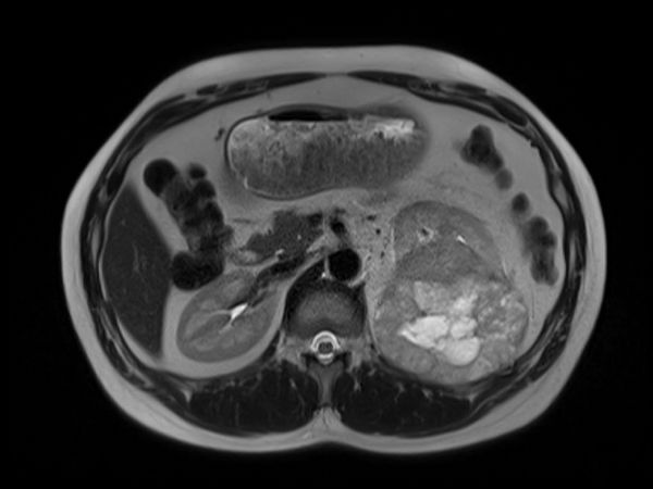
Resim 1
Figure 1
)
Sol bobrek ust-orta polde 10x8 cm boyutunda içinde kistik komponenti bulunan ve nekroze alanlar iceren, belirgin kontrast tutulumu gosteren T1 agırlıklı goruntude hipointens hipointens T2 de hafif hiperintens ozellikte solid kitle
MRI of abdomen was reported as a 10x8 cm solid mass of the left kidney enhancing contrast with necrosis and cystic components
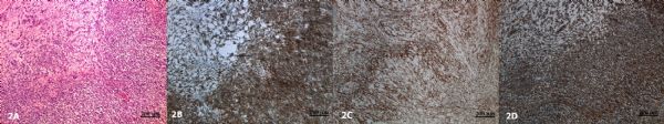
Resim 2
Figure 2
)
2A: Kit sitoplazmali kucuk yuvarlak hucrelerden olusan blastemal komponent ve stromal komponent (HEX 100).
2B: Tumor hucrelerinde izlenen diffuz CD56 immunopozitivitesi (X100).
2C: Tumor hucrelerinde izlenen diffuz WT1 immunopozitivitesi (X100).
2D: Tumor hucrelerinde izlenen diffuz Vimentin immunopozitivitesi (X100).
2B: Tumor hucrelerinde izlenen diffuz CD56 immunopozitivitesi (X100).
2C: Tumor hucrelerinde izlenen diffuz WT1 immunopozitivitesi (X100).
2D: Tumor hucrelerinde izlenen diffuz Vimentin immunopozitivitesi (X100).
2A: The blastemal component, consist of small round cells with scant cytoplasm and stromal component (H&EX 100).
2B: The tumor cells show immunoreactivity for CD 56 (X100).
2C: The tumor cells show immunoreactivity for WT1 (X100).
2D: The tumor cells show immunoreactivity for Vimentin (X100).
2B: The tumor cells show immunoreactivity for CD 56 (X100).
2C: The tumor cells show immunoreactivity for WT1 (X100).
2D: The tumor cells show immunoreactivity for Vimentin (X100).
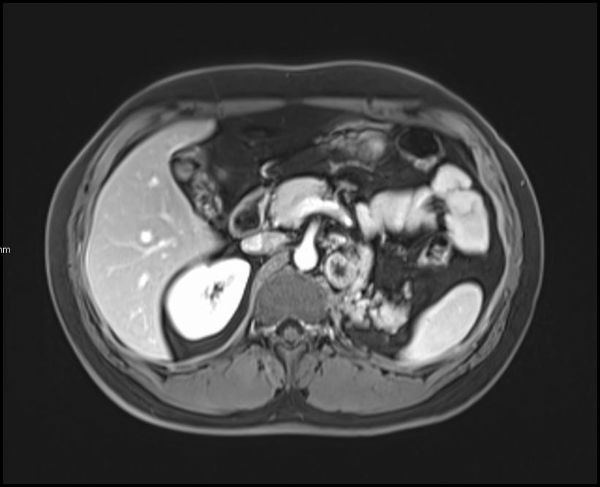
Resim 3
Figure 3
)
Operasyon ve KT sonrasi sol bobrek lojunda ve batin icinde multipl metastazla uyumlu lenf nodu
Multiple masses at the nephrectomy region that the biggest one measured 18x18 mm and a 8 mm nodle at the third month of follow up