[P-141]
Oturum adı: POSTER SESSION 3 | Oturum salonu: POSTER AREA | Oturum tarihi: 17 Ekim 2014 | Oturum saati: 14:00 - 19:00DJ Stent Takılmasının Nadir görülen Bir Komplikasyonu
Mehmet Mazhar Utanğaç, Ahmet Ali Sancaktutar, Mehmet Nuri Bodakçı, Mansur Dağgülli, Onur Dede, Necmettin Penbegül, Namık Kemal Hatipoğlu, Haluk Söylemez, Yaşar BozkurtAMAÇ: Endoürolojik girişimlerin hızla artmasıyla DJ stent kullanımı da giderek artmaktadır. Sık kullanılmalarının bir sonucu olarak DJ stente bağlı komplikasyonlar da artmıştır. Bunlardan biri de DJ kateterin takılırken guide’ın çekilmesi sırasında düğümlenmesidir. Burada üreter taşı nedeniyle üreterorenoskopi işlemi sırasında DJ stent takılmaya çalışılırken düğümlenen bir olguyu sunmayı amaçladık.
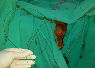
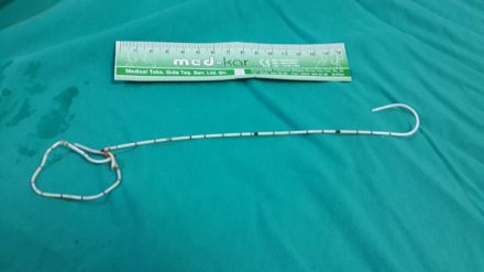
CASE: 59 Yaşında erkek hasta sol yan ağrısı nedeni ile dış merkezde yapılan görüntüleme sonucunda sol üreter taşı saptanması üzerine hastaya endoskopik taş kırma üreter ve sonrasında DJ stent takılması planlanmış. Hastaya DJ kateter takılırken DJ kateterinin içinden gönderilen guide’ın çekimi sırasında takılıp gelmemesi üzerine çekilen skopi görüntülenmesinde DJ kateter ile beraber içinden gönderilen guide’ında üst üreter içinde düğümlendiği görülmüş. Bunun üzerine üreter daha fazla zorlanmadan hasta kliniğimize yönlendirilmişti. Hasta kliniğimize başvurduğunda DJ stentin guide’ı üretral meadan dışarı çıkmış haldeydi (Resim 1). Çekilen direk grafide düğümlenmiş şekilde DJ stentin üst üreterde olduğu gözlendi (resim2). Hastaya üretroskop ile girildi. Üreter içinde düğümlenmiş olan DJ stent ve guide görüldü. Bunun üzerine lazer ile DJ stent ve guide düğümlendiği yerden kesilerek yabancı cisim forsepsi ile dışarı alındı (resim 3). Hastaya retrograde opak madde verilerek üreterin intakt olduğu gözlendi. Yeniden DJ stent yerleştirildi. Post operatif çekilen direk grafide DJ stentin yerinde olduğu gözlendi. Bir ay sonra DJ stent çekimi yapılmak üzere hasta taburcu edildi.
SONUÇ: Üroloji pratiğinde sıkça kullanılan DJ stent takılması çok nadir de olsa komplikasyon ile karşımıza çıkabilmektedir. Üreter içerisinde düğümlenmiş halde bulunan DJ stentin lazer kullanılarak fragmante hale getirilip dışarı alınması bir yöntem olarak kabul edilebilir.
A Rarely Seen Complication Of DJ Stent İmplantation
Mehmet Mazhar Utanğaç, Ahmet Ali Sancaktutar, Mehmet Nuri Bodakçı, Mansur Dağgülli, Onur Dede, Necmettin Penbegül, Namık Kemal Hatipoğlu, Haluk Söylemez, Yaşar BozkurtOBJECTIVE: With rapid increase in endourological interventions DJ stents are also used more frequently. As a result of their frequent use, DJ stent-related complications have also increased in number. One of them is knotted DJ catheter upon withdrawal of the guidewire during implantation of the DJ catheter. Herein, we aimed to present a case where DJ stent was knotted during ureterorenoscopy procedure performed for the extraction of a ureteral stone.
CASE: A 59-year-old male patient had been referred to an external center with complaints of left flank pain, and radiological images obtained had revealed the presence of a left ureteral stone which necessitated planing of endoscopic stone fragmentation, and subsequent DJ stent implantation. During withdrawal of the guidewire which had been sent through DJ catheter, guidewire resisted its removal. Fluoroscopic examination had revealed the presence of a knotted DJ catheter, and its guidewire within the upper ureter. Then without any further forceful attempt at withdrawal, the patient was referred to our clinic. At admission guidewire of the DJ stent was protruding from urethral meatus (Figure 1). KUB grapy demonstrated a knotted DJ stent within the upper ureteral segment (Figure 2). On ureteroscopic examination, knotted DJ stent with its guide wire was seen. Then using laser, the knotted segment of DJ stent, and guide wire were cut, and taken out with a foreign substance forceps. (Figure 3). Opaque material was instilled retrogradely through ureter, and its intactness was confirmed. A new DJ stent was inserted. On postoperative KUB grapy the proper position of DJ stent was ensured. The patient was discharged, and a return visit was planned to remove DJ stent one month later.
CONCLUSION: In urology practice frequently used DJ stent implantation though very rarely can cause complications. Removal of the knotted DJ stent in the ureter after its fragmentation using laser can be considered as a useful method in such complicated cases.
Resim 1: Üretral meadan dışarıda görülen DJ stentin guide’ı
Figure 1: Guidewire of the DJ stent is seen protruding from urethral meatus
)
Resim 2: Direk grafide düğümlenmiş şekilde DJ stent
Figure 2: Kidney ureter bladder grapy demonstrated a knotted DJ stent

)
Resim 3: Dışarı alınan DJ stent
Figure 3: Taken out DJ stent
)