[P-002]
Oturum adı: POSTER SESSION 1 | Oturum salonu: POSTER AREA | Oturum tarihi: 16 Ekim 2014 | Oturum saati: 14:00 - 19:00Genç bir erkekte üretral obstrüksiyonun beklenmedik bir nedeni: Benign prostat hiperplazisi
Akif Koç1, Erhan Sarı1, Erdoğan Bülbül2, Ahmet Küçükyangöz1, Bahar Yanık22Balıkesir Üniversitesi, Radyoloji Anabilim Dalı, Balıkesir
AMAÇ: Bir genç erkekte infravezikal obstrüksiyona neden olan benign prostat büyümesi (BPH) olgusu sunuldu.
OLGU: 17 yaşında erkek hasta idrar yapamama şikayet ile dış merkeze başvurmuş. Yapılan sistoskopisinde prostat sol lobunun prostatik üretraya doğru hipertrofik olduğu gözlenmiş. Başka patoloji tespit edilmeyen hastaya 16 F foley sonda konularak kliniğimize yönlendirilmiş. Özgeçmiş ve soy geçmişinde özellik bulunmayan hastanın laboratuar bulguları; üre:22 mg/dl, kreatinin:0,6 mg/dl, glukoz:98 mg/dl, wbc:5,2 103/µL, Hb:10,5 g/dL, crp:0,7 mg/L, TİT: 1018/ph:5,5/28 ertrosit/5 lökosit şeklinde idi.
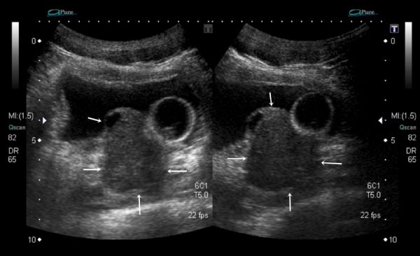
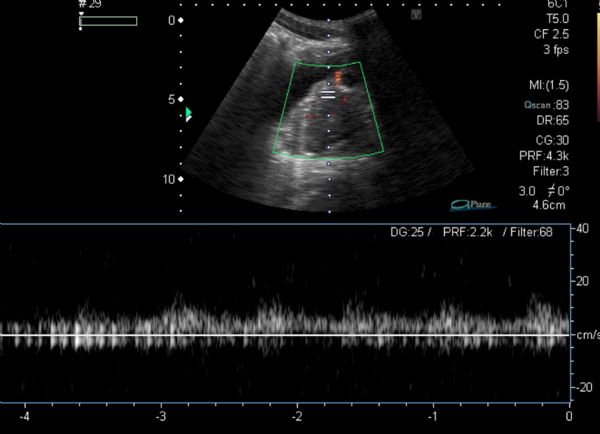
Suprapubik abdominal ultrasonografi (USG) incelemesinde prostat glandı boyutları artmış olduğu gözlendi. Prostat içerisinde milimetrik kistik komponentler bulunduran orta hattın hafif sağında yerleşen, boyutları yaklaşık 42x38x39 mm ölçülen hipoekoik solid lezyon saptandı. Lezyon içerisinde doppler USG incelemede arteriel akım örnekleri belirlendi. Prostat gland kaynaklı yer kaplayıcı oluşum olarak değerlendirildi. Tanımlanan lezyon mesane tabanına bası oluşturduğu gözlendi. (Resim 1,2)
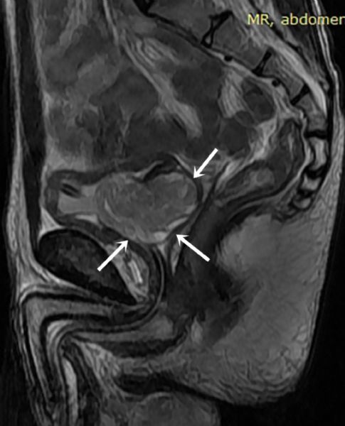
Yapılan tüm abdomen manyetik rezonans görüntülemede (MRG) prostat glandı sağ kesiminde, sinyal intensitesi T1A sekansta komşu kas dokular ile izointens, T2A sekansta hiperintens kitle görüldü. Boyutları 4,5*2,5 cm ölçüldü. İçerisinde fokal hiperintens kistik komponentler ve hafif kontrast tutulumu izlendi. Prostat gland posterior konturunda lobülasyon, rektovezikal resese protrüzyon belirlendi. Lezyon öncelikle prostat kaynaklı tümör (stromal tümör?) lehine rapor edildi. (Resim 3)
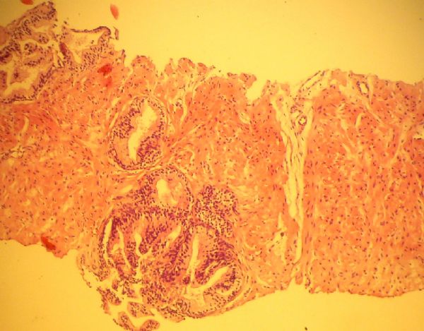
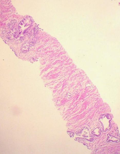
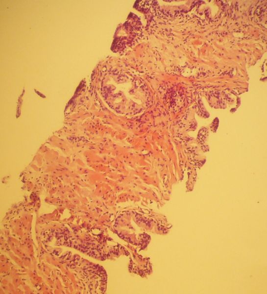
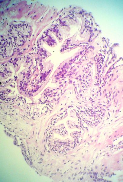
Takiben hastaya TRUS eşliğinde prostat biyopsisi yapıldı. Patolojisi Düzenli asiner yapılardan, düz kas demetlerinden, bağ dokusundan ibaret prostat dokusu izlenmektedir. Malignite izlenmemiştir. şeklinde rapor edildi (Resim 4,5,6,7)
Operasyon planlanan hasta ileri merkeze yönlendirildi.
SONUÇ: Genellikle ileri yaştaki erkek hastalarda infravezikal obstrüksiyona neden olan BPH çok nadir olmakla birlikte infravezikal obstrüksiyon şüphesi olan genç yaştaki erkeklerde de etiyolojide akla gelmelidir.
An unusual cause of urethral obstruction in a boy: benign prostate hiperplasia
Akif Koç1, Erhan Sarı1, Erdoğan Bülbül2, Ahmet Küçükyangöz1, Bahar Yanık22Department of Radiology, Balıkesir University, Balıkesir, Turkey
OBJECTIVE: A case of benign prostatic enlargement which causes infravesical obstruction in a young boy was presented.
CASE: A 17 year old male patient had admitted to the outer center with the complaint of inability to urinate. In his cystoscopy, the prostate hypertrophy of the left lobe into the prostatic urethra was observed. Patient who did not identify another pathology, was placed16 F Foley catheter and referred to our clinic. There was not any significant feature in his personal and family histories. Laboratory findings was determined as; urea: 22 mg / dL, creatinine: 0.6 mg / dL, glucose: 98 mg / dl, WBC: 5.2 103/µl, hemoglobin: 10.5 g / dL, CRP: 0.7 mg / L, TIT: 1018/ph: 5.5 / 28 erythrocyte / 5 leukocytes.
Suprapubik abdominal ultrasonography (USG) revealed increased size of the prostate gland.
A hypoechoic solid mass which had approximately 42x38x39mm dimensions and containing cystic components was observed at the right side of the prostate gland. The arterial flow samples in the lesion was taken with doppler USG examination. The mass was considered as a space-occupying formation originating from the prostate gland. The defined lesion constituted press to the base of the bladder. (Fig. 1,2)
In magnetic resonance imaging (MRI), a mass detected at the right side of prostate gland with T1W isointense with adjacent muscles and T2W hyperintense signal. Dimentions was measured 4,5x 2,5cm. The mass was containing focal hyperintense cystic components and was minimally enhancing. Lobulation and protrusion to the rectovesical recess was identified at the posterior prostate contour. Lesion was initially reported as a prostate gland originating mass ( stromal tumour?). (Fig. 3)
Subsequently, the patient underwent TRUS-guided prostate biopsy. Pathologic examination result was reported as " The prostate tissue which consisted of regular acinar structures, smooth muscle bundles and connective tissue was observed. Malignancy wasn't observed. " (Fig. 4,5,6,7)
Operation planned patient was reffered to an advenced center.
RESULTS: BPH which often causes infravesical obstruction in elderly male patients, also should be considered as an etiologic reason in young male patients who are suspected to infravesical obstruction.
Resim 1
Figure 1
Suprapubik abdominal ultrasonografi görüntüsünde prostat içerisinde kistik komponent bulunduran hipoekoik yer kaplayıcı lezyon izlenmiştir.
Hypoechoic solid, space-occupaying lesion containing cystic component was seen at suprapubic abdominal ultrasonography image.
Resim 2
Figure 2
Renkli doppler ultrasonografi görüntüsünde kitle içerisinde düşük direçli arteriyel akım belirlenmiştir.
Low resistance arterial flow was detected within the mass in his collored doppler ultrasonography.
Resim 3
Figure 3
Sagital yağ baskılı manyetik rezonans görüntüde prostat glandı içerisinde kistik komponentler bulunduran, posterior protrüzyon gösteren kitle belirlenmiştir.
In sagital fat supression magnetic resonance image posterior protruding mass with cystic components was detected.
Resim 4
Figure 4
Düzenli asiner yapı ve düz kas dokusu HE 12x20
Regular acinar structure and smooth muscle tissue HE 12x20
Resim 5
Figure 5
